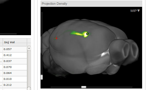
 I am currently modeling the projections from CA1 and Subiculum to Entorhinal Cortex using PAM. In the 3D-model, it is not so obvious how they should look like. The best, I could find is the collection of tracer studies that is available at the Allen Brain Atlas. For over 1000 injection sites you can analyse where in 3d-space the projections of the neurons are. In the example above you see, how CA1 neurons (green dot) project to the Entorhinal cortex (red cross). The green trace connecting the dot with the cross is almost invisible.
Martin Pyka
Neuroscience, Data Analysis, Art, Stuff