
This is great scientific nonsense.
Origin of the Novel Species Noodleous doubleous
Reply
This is great scientific nonsense.
Recent reports have shown that theta oscillations are in fact traveling waves along the septo-temporal axis of the hippocampus (at least in rats).
Lubenov and Siapas, 2009, Hippocampal theta oscillations are travelling waves
Patel et al., 2012, Traveling Theta Waves along the Entire Septotemporal Axis of the Hippocampus
Here is a little animation based on my 3d model of the rat hippocampus to show how the traveling waves might look like in CA1.
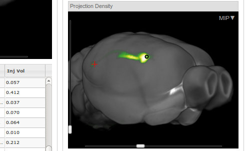 I am currently modeling the projections from CA1 and Subiculum to Entorhinal Cortex using PAM. In the 3D-model, it is not so obvious how they should look like. The best, I could find is the collection of tracer studies that is available at the Allen Brain Atlas. For over 1000 injection sites you can analyse where in 3d-space the projections of the neurons are. In the example above you see, how CA1 neurons (green dot) project to the Entorhinal cortex (red cross). The green trace connecting the dot with the cross is almost invisible.
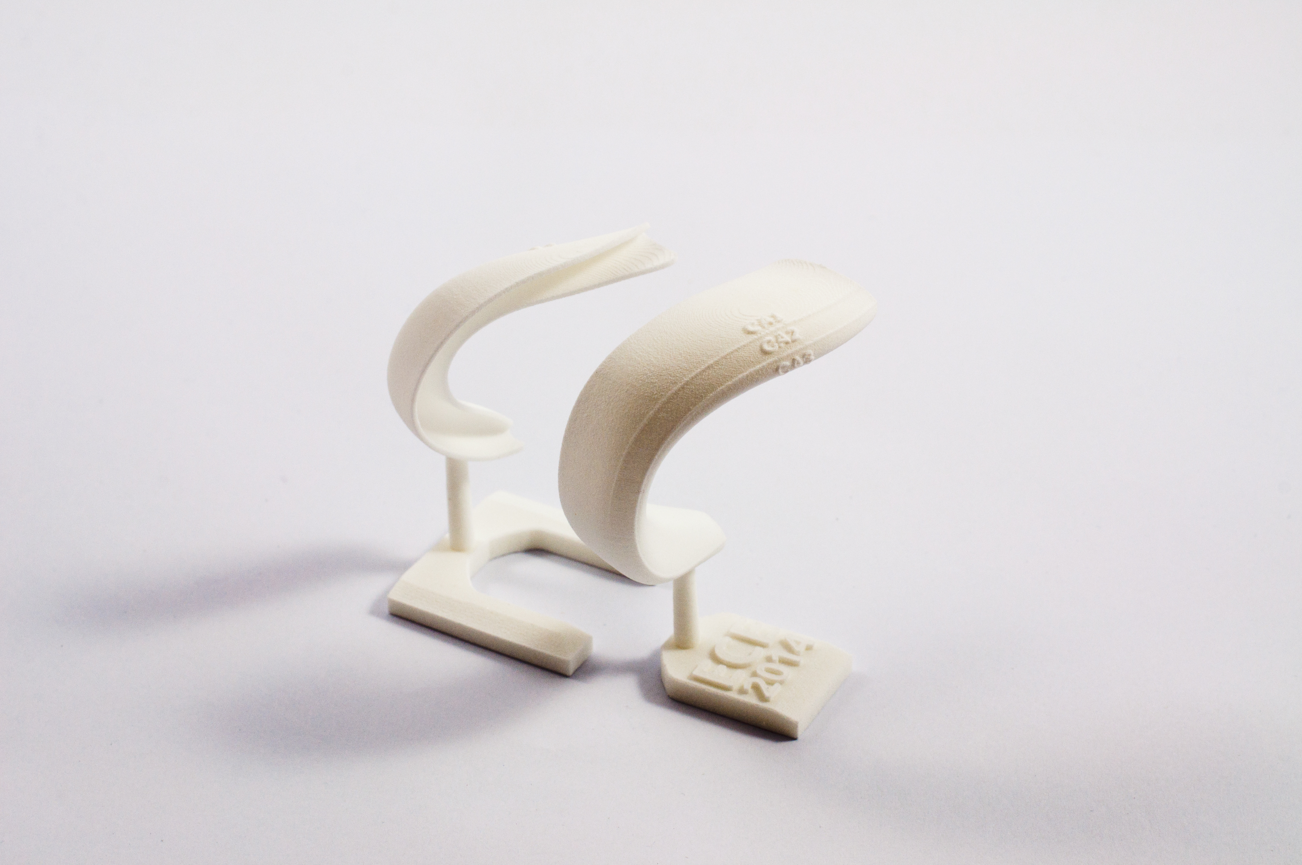
Recently, we had the Memory and Mind workshop of the European Cluster of Excellence in our lab. On short notice, the organizers of this workshop approached me to ask whether I could organize 54 3d-printed models of the rat hippocampus for the participants. The models need to be printed within the next 7 days in order to give them to the participants.
It turned out, that most 3d printing services (and we needed one with selective laser sintering) simply cannot deliver within such a short time. Fortunately, I found trinckle, a startup in Berlin, who managed to produce and deliver 54 models of the rat hippocampus right on time to hand them out to the participants of the workshop at the last day. They now published a nice posting about the hippocampus and the 3d-model (in German). Thank you!
NEURO tv is a monthly online conversation between neuroscientists, psychologists and philosophers who try to understand the brain and the mind.
Jep, and this is great! Jean-Francois Gariépy uploaded already 11 episodes of his Neuro.TV series, each of them roughly an hour long. He organizes online video meetings with researchers discussing latest opinions and research directions in neurosience.
Scaling Brain Size, Keeping Timing: Evolutionary Preservation of Brain Rhythms by György Buzsáki, Nikos Logothetis and Wolf Singer
In this paper, they report changes of frequency of several rhythms as a function of brain weight. What I found interesting was Fig. 2 showing that most rhythms are invariant to brain weight (or equally size) except theta, suggesting that spatial properties of the hippocampus (e.g. its overall size) indeed constrain the range in which theta rhythm emerges.
I am proud to announce the start of a new long term project that begins with a paper that came out recently.
PAM is the foundation for studying generic and specific coding principles of neural networks. The idea is to create artificial neural networks whose connectivity properties and connection lengths result from reconstructed anatomical data. Using a 3d environment to model and describe real neural networks, we can describe complex relationships between layers of neurons. With PAM, we can account for connectivity properties that are influenced by global and local anatomical axes, non-linear relationships between distances of neurons and their connection lengths, and we can incorporate many experimental data, like images from tracer studies, directly into our model.
In the long run, I hope that PAM can help to understand how the structure but also how self-organizing principles (like diverse forms of plasticity) contribute to the topology and thereby also to the function of the network. In particular, we are interested in the functioning of the hippocampus.
The project website is hosted on Bitbucket Github.
Updated on 26.05.2015:
Replaced link to Bitbuckt by link to Github.
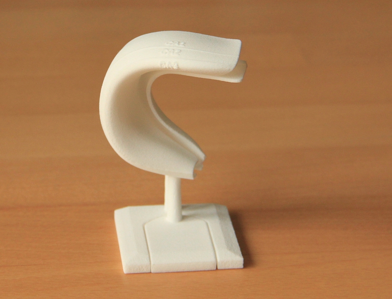 If you, like me, work on the hippocampus, you know that it is incredibly hard to understand how the hippocampal formation actually looks like in 3D. Dentate Gyrus and CA3-CA1 lie in the rat along the dorsal-ventral axis but also traverse the medial-lateral and anterior-posterior axis.
Now you can order a 3d print of the hippocampus at Shapeways which helps a lot to understand how the neural layers look like and how they are interwoven.
This TED talks shows some impressive visualizations of molecular reactions. In particular the emergence of this football-like shape (at around 1:16) is astonishing.
The software they use, called molecular flipbook, has been developed by a team around Janet Iwasa and is based on Blender. This is another great example of Blender becoming more and more a helpful tool for scientists.

The Pigeon Brain Atlas
Recently, a 3d atlas of the ascending sensory and the descending motor systems in the pigeon brain has been presented, that is freely available. This is one important step towards a database that allows comparisons of network structures and their functions across species.
